The main danger of infectious diseases in cows is that they quickly spread to the entire livestock of the farm. In addition, many of them have a high mortality rate. And one of the champions in this case is rinderpest. This is an extremely contagious disease, which in almost 100% of cases ends in death. That is why it is important to know what this disease is and how to protect the herd from it.
Plague in cattle
What is rinderpest?
In addition to cows, rinderpest, also referred to as Pestis bovina, spreads to goats and sheep. Among wild animals, deer, antelopes, buffaloes, camels and many other ungulates are susceptible to it. This disease is viral in nature. It affects the mucous membranes of the digestive tract, intestines, respiratory tract, and also the skin. In this case, the affected area becomes sharply inflamed and quickly undergoes necrotic processes, on the basis of which blood infection and death of the animal occur.
History
According to the international classification, this disease belongs to group A (especially dangerous infectious diseases). It is worth noting that it has been known for a long time. The first information about this disease is found in ancient Roman written sources, which were created in the XNUMXst century AD. Later, by the beginning of the XNUMXth century, a strong epidemic was traced in the countries of Asia.
The most widespread spread of rinderpest in Europe falls on the 1841th century. From Holland, Germany and England, it very quickly spread to all European countries. At the same time, the disease caused enormous damage to the economy, and agriculture in particular, for almost two centuries. In XNUMX, infected animals came to Egypt, from where the epidemic quickly spread to all countries of the mainland, destroying livestock.
Since the end of the 1924th century, an active fight against the disease began. In 1928, they were able to liquidate it throughout Australia. By 2011 rinderpest was completely defeated in the USSR. In XNUMX, the UN announced worldwide that the last foci of this disease were found and destroyed in African countries.
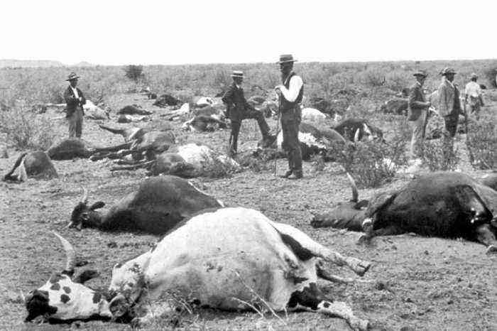
Plague in Africa
Causative agent
The causative agent of the disease is a special virus, which is one of the paramyxaviruses and contains its own RNA. The species that causes distemper in cattle is similar in its specificity to the distemper virus in dogs and the causative agent of measles in humans. When it enters the bloodstream, the infection immediately spreads through the bloodstream. At the same time, the largest accumulations of pathogen bodies can be traced in the lungs, lymph nodes and kidneys. But, in the end, it affects all organs and tissues of the animal.
The infection reacts to the negative influences of the external environment in different ways. The most destructive for her are:
- direct sunlight. Kill the infection within 1-5 hours;
- sub-zero temperatures. Virus inactivation occurs within 30 days;
- temperature over 60 degrees. Kills the pathogen in 15-20 minutes;
- sodium hydroxide. A 2-minute exposure is enough;
- in manure, the virus remains active for one day;
- at room temperature can live more than 5 days.
Causes of appearance
Rinderpest spreads to most artiodactyl domestic and wild animals. It is most dangerous for young individuals, over the age of one year. The main source of transmission of the virus is an infected animal. It releases pathogens into the environment along with:
- nasal secretions;
- fecal masses;
- urine;
- blood;
- milk;
- mucus from the conjunctiva;
- secretions from the genitals.
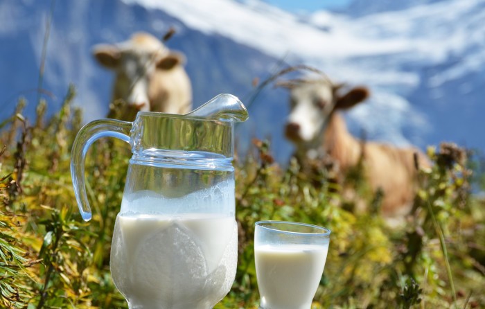
Pathogens are released into the environment along with milk
As a rule, in all these substances the virus appears on the 3rd-8th day of illness. In the mucous secretions of the genital organs, even after complete recovery, the infection persists and can be transmitted to other animals within 3 weeks.
There are many ways of transmitting the virus to healthy animals. Among them the main ones are:
- Air. The pathogen enters the cow’s respiratory tract through the air, after which the infection spreads. Contribute to this form of distribution of crowded content and weak immunity of livestock.
- Alimentary (oral-fecal). Particles from the secretions of sick animals, in which the virus is located, enter the feed and water. When they are consumed, they become infected. Especially often this method of transmission of the disease occurs in farms that do not comply with sanitary standards, there is no regular cleaning of the premises and periodic disinfection.
- Mechanical. The source of the spread of the disease in this case may be the carcasses of animals that have died from the plague. Birds and dogs and pigs feed on these bodies, which, upon contact with the cow, transmit the infection to her.
The virus can also be transmitted through the clothing and tools of service personnel. It was found that blood-sucking insects, when absorbing blood for 15 minutes, can also be carriers. But cases of transmission of the pathogen through ticks, horseflies, mosquitoes have not been identified. For a long time, the pathogen can live in the skin, meat, and horns of dead livestock.
Important! When released into livestock farms, the disease progresses at any time of the year. It spreads especially intensively on farms with a high concentration of animals.
Symptoms
The incubation period for the virus in most cases of infection is from 3 days to a week. Less often, the terms can reach up to 15-17 days. At the end of the incubation period, the disease begins to actively develop on the walls of the mucous membranes of the intestines and respiratory tract. Soon, the infection corrodes the walls of the mucosa and blood vessels, entering the blood. At the site of the lesion, necrotic processes quickly appear and ulcers appear, which are covered from above with a liquefied layer of dead epithelium.
Symptoms of the disease
After entering the blood, the virus through the circulatory system reaches the lymph nodes, lungs, spinal cord, kidneys, causing pathological changes in them. The development of the virus quickly inactivates the body’s immune system.
The further course of the disease involves the passage of three main stages.
The first stage
It is also called feverish. It develops immediately after the end of the incubation period. This stage is accompanied by such clinical signs:
- a sharp increase in the cow’s temperature to 41-42 degrees;
- more agitated behavior;
- rapid heartbeat and breathing;
- refusal of food, in parallel with which excessive fluid intake can be traced;
- the skin on the nose becomes dry;
- the mucous membrane of the eye and throat become red, the initial stages of the inflammatory process are visible;
- increased sensitivity of the animal to bright light;
- wool becomes brittle, loses its luster, constantly disheveled.
Usually the duration of this stage does not exceed 2-3 days.
The second stage
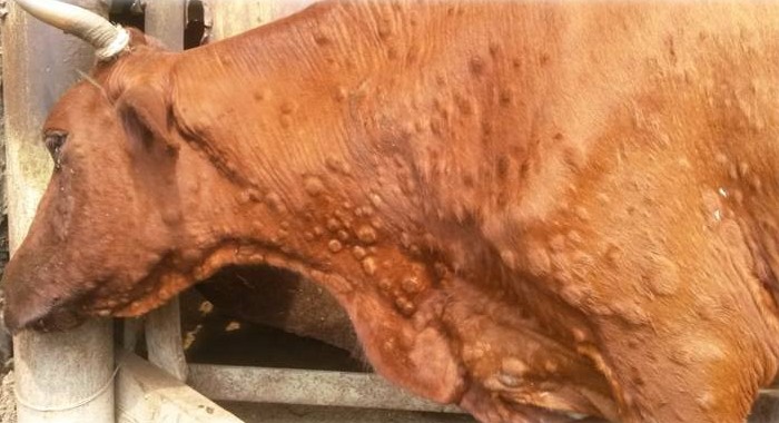
After 3 days of the primary development of the disease, necrosis of the tissues of the mucous membrane begins to appear actively. Moreover, ulcers and erosions are visible on the conjunctiva, in the oral and nasal cavities. This stage of the disease is accompanied by the following symptoms:
- Serous conjunctivitis develops rapidly, which passes from serous to purulent form within a day or two.
- A purulent discharge flows from the nose, which dries up in the form of scabs under the nostrils. Pus accumulates in large quantities in the nasal cavity itself.
- The mucous membrane of the eyes and nose is red and swollen.
- The animal constantly sneezes, uneasily shakes its head, and can shift from foot to foot.
- The mucous membrane of the mouth, palate, lips, cheeks from the inside are covered with small dots of gray or yellowish color. These are the initial foci of tissue necrosis.
- Increased salivation. The saliva has a foamy appearance and is speckled with inclusions of blood.
- Swallowing is accompanied by severe pain.
- Grayish or yellow foci of erosion also appear on the vulva. Exudate with blood impurities periodically comes out of the genitals.
This phase of the disease in pregnant individuals certainly leads to fetal death and abortion. It usually ends with a gradual decrease in temperature.
Examining a pregnant cow
The third stage
This stage of the course of the disease is characterized by a serious lesion of the mucosa of the gastrointestinal tract. It traces:
- Normalization of temperature or its fall below the natural mark;
- Severe diarrhea, which with development turns into an involuntary excretion of feces;
- In the feces, impurities of blood, mucus, particles of dead tissues of the intestinal walls are traced;
- The mucous membrane of the anus is strongly protruded and painted in a dark red color;
- The animal feels pain during bowel movements and just standing still. Therefore, in order to weaken it, the back is arched;
- Breathing is heavy and very rapid.
With such disorders of the digestive system, all water quickly leaves the body, and the animal does not have time to replenish its reserves. Against the background of this phenomenon, a rapid loss of strength and weight is manifested. An animal can lose up to 30% or more of its original weight in a matter of hours. Death usually occurs by day 9 from the end of the incubation period.
It is worth noting that in some cases a latent course of the disease is also possible. Symptoms are practically not expressed, mucous membranes are not damaged. Often this form of plague ends in the recovery of the animal. In this case, it acquires a strong immunity to re-infection for a period of 5 years or more.
Due to the severely weakened immune system, other diseases can develop against its background. This aggravates the condition of the cattle and complicates the diagnosis, because characteristic secondary features are also added to the primary signs.
Diagnostics
Pestis bovina is similar to a number of other infectious diseases in a number of clinical ways. Therefore, it is not possible to make an accurate diagnosis only on the symptoms that appear in a cow. To verify the preliminary conclusions, laboratory diagnostics and pathoanatomical examination are carried out.

Laboratory diagnosis
For laboratory study, prescapular and other lymph nodes, particles of the spleen, liver or lungs of dead animals are taken as material. Blood is taken from living individuals for analysis. Diagnostics in the laboratory is carried out in three ways:
- Identification of the pathogen in the blood using a binding reaction or enzyme immunoassay.
- Determination of specific antibodies in blood and other tissues.
- Detection of changes in the structure of cells and the composition of the cytoplasm, which indicate the presence of the virus.
It should be noted that tissues from the bodies of dead cattle should be taken no later than 5-6 hours after their death. The material is placed in a sealed container and sent to the laboratory.
Pathological anatomical examination of the presence of rinderpest may indicate serious changes in internal organs. The organs of the digestive system are characterized by:
- cheesy plaque on the mucous membranes;
- the mouth, throat, lips and cheeks of the animal are covered with gray nodules or ulcers;
- the lymph nodes of the mesentery of the intestine are greatly enlarged and inflamed;
- the intestinal mucosa is covered with pinpoint ulcers and hemorrhage;
- the thickness of the walls of the small intestine is greatly increased.
The lungs of the cow are inflamed, all the lymph nodes of the body are enlarged and also suggest signs of inflammation. At autopsy in the abdominal cavity of the carcass is a small amount of cloudy darkish liquid.
Treatment
It should be noted that the fight against rinderpest is strictly regulated by international veterinary legislation. In accordance with it, any measures in the direction of the treatment of the disease are prohibited. Moreover, an effective therapeutic method in this direction has not been identified.
If, as a result of the diagnosis, plague was detected in livestock, all infected animals are slaughtered as soon as possible. Their corpses are immediately burned. The only measure to combat such a disease is high-quality prevention.
Prevention
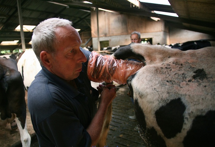
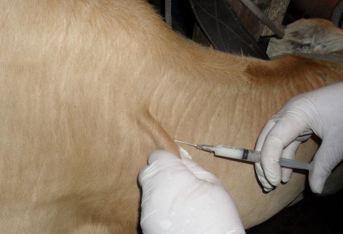
The main preventive measure against the occurrence and spread of the disease is the periodic vaccination of young and adult livestock of the farm. To do this, drugs based on live and deactivated cultures of the virus are used. The injection of such a vaccine is carried out subcutaneously at a dose of 1 ml. As a result, antibodies are produced in the body that can resist infection from outside. The process of developing a protective mechanism lasts for 3 days. After that, the cattle develops immunity, which lasts for the next 3 years.
Cow vaccination
If individuals with rinderpest are detected in a certain farm, this farm, and the entire settlement in which it is located, are transferred to a quarantine regime. It includes the following points:
- Ban on the export of animals, meat and dairy products, animal skins, fodder stocks. The introduction of new animals into the quarantine area is also prohibited.
- The movement of people and the departure of vehicles outside the settlement is prohibited. For this purpose, police posts with a round-the-clock watch are established at all entrances and exits.
- Pasture maintenance and general watering are completely excluded.
- A dedicated team of veterinarians inspect the livestock twice a day. If signs of illness are detected or the temperature does not drop within 2-3 days, the animals are killed. At the same time, the carcasses of slaughtered livestock are burned immediately.
- All livestock premises are cleaned every day. All withdrawn manure is also burned. After cleaning, the barn is disinfected with caustic soda at a dosage of 1,5 liters per square meter of area or other solutions.
- All cattle in the village are vaccinated.
If all the measures taken do not bring improvements, the district administration may decide to slaughter the entire livestock in the village. After that, all the premises where he was kept are cleaned and disinfected.
If the quarantine gave the desired effect and the disease was eliminated, it is maintained for another 21 days. After that, several heads of young animals that have not been vaccinated against the plague are launched into an empty barn for testing. If no signs of the disease are detected within a month, breeders are allowed to launch new animals. The import of new livestock is carried out with mandatory isolation, the period of which is 15 days. It must be vaccinated before import.
Attention! It is allowed to take animals to other settlements only after 6 months. All animals on the affected farm are vaccinated every year for the next three years. At the same time, the farm is subject to regular inspections by the district sanitary department.
Conclusion
Rinderpest is now considered a completely eradicated disease. But, given the danger of this disease and the extent of the damage that it can cause, it is impossible to relax attention on this issue. Therefore, every breeder is obliged to know the symptoms of the disease in order to prevent its spread in time.


