Among various parasitic diseases of cows, dicroceliasis of cattle stands out in a separate category. What makes this disease special is the fact that it also affects small livestock, dogs, pigs, horses, wild ruminants and humans. Accordingly, the appearance of an outbreak on a single farm threatens a pandemic for all its inhabitants. At the same time, the danger of the disease lies in the fact that in some cases it can end in severe exhaustion and death of the sick individual.
healthy cow
Causative agent
Dicrocoeliosis is a chronic disease that is accompanied by a decrease in the productivity of individuals, disruption of the digestive tract, and general exhaustion. It occurs in most domestic and wild animals, as well as in humans. In ruminants, the disease is most severe and often ends in the death of the infected individual. The causative agent of the disease is a special type of trematode – Dicrocoelium lanceatum.
Description
This helminth affects mainly the liver and gallbladder. The trematode belongs to the category of lanceolate. The body of the worm tapers towards the ends, has a dark gray color, and its length is from 5 to 15 mm. There are suckers on the body, with the help of which the parasite is fixed in the bile ducts. Behind the ventral sucker are the testes, near the front is the genital opening.
The parasite reproduces by laying eggs. They are brown in color and asymmetrical in shape. Also, the features of the eggs of this trematode include a thick shell that protects the larva (miracidia) from harmful external influences.
The biological cycle of the pathogen
Dicrocoelium lanceatum requires intermediate hosts for a complete development cycle. They are ants and mollusks (10 species living on land). The whole process of helminth development is as follows:
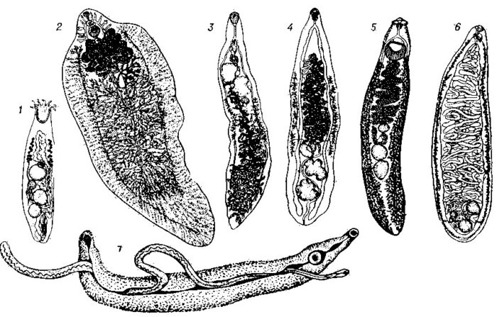
Full cycle of Dicrocoelium lanceatum
- Adult dicrocelia, parasitizing in the bile ducts of the liver, lay eggs, which, along with the flow of bile, enter the intestine.
- Here, bile is mixed with feces, and during defecation, the eggs enter the external environment.
- Miracidia, enclosed in a shell, are swallowed by freshwater mollusks in the external environment.
- In their body, the larva is released from the egg and moves to the midgut of the snail, where it gradually passes into the sporocyst stage. Inside such a formation, a number of daughter sporocysts are formed, and after their full maturation, the maternal one completely disappears.
- From daughter cysts, another stage of larvae is formed – cercariae. Such an organism travels to the lungs of the intermediate host, where the larvae again transform into a cyst form. They are collected in mucous lumps, each of which contains 100-300 dicrocelia.
- Formed mucous lumps through the respiratory cavity enter the external environment, where they are fixed with the help of mucus on surrounding objects and plants.
- In the future, such lumps are collected and eaten by ants. At the same time, getting into the belly of the insect, the larva is released from the shell and develops to the stage of metacercaria.
- Infection of a cow occurs while eating grass with ants, which turn out to be carriers of the developing helminth.
In general, the full development cycle of the larva of dicrocelia up to entering the body of the final host takes 3,5-4,5 months. Another 1,5-3 months is the final maturation in the body of a cow or other animal. The invasion can last for several years, negatively affecting the growth and development of livestock.
Resistance to external influences
In the natural environment, miracidia in the shell are extremely stable. The larva is able to maintain its viability under such influences:
- at a temperature of 50 degrees, it survives for a day;
- lowering to -50 degrees transfers without damage;
- weekly drying in direct sunlight at a temperature of 20 degrees also does not affect the viability of the larva;
- in the metacercariae stage, the helminth can safely winter in the body of ants.
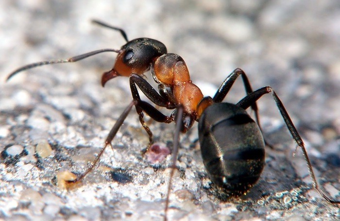
In the metacercaria stage, the helminth can overwinter in the body of an ant.
From the respiratory tract of mollusks, larvae are most often released into the external environment after rain. At this time, the air humidity rises above 67%, and the temperature drops. Such conditions contribute to increased activity of snails, and parasites, respectively, are able to spread more actively.
Infection of cows on pastures, as a rule, takes place during the first pastures. After 1,5-2 months, the feces of animals already contain trematode eggs. The scale of infection is especially high in the spring-summer period. Outbreaks of the disease can be traced in various countries. But in regions with a warm climate, epidemics are more widespread and occur much more frequently.
Symptoms
The degree of manifestation of clinical signs of dicroceliosis largely depends on the number of helminths parasitizing in the body, as well as on the general physical condition of the animal. With minimal invasion, the symptoms of the disease are practically not manifested. In this case, trematodes can freely live in the liver for several years.
If the degree of infection is high, the disease may be accompanied by such manifestations:
- anemia;
- gradual weight loss of the animal;
- yellowing of mucous membranes;
- violation of the digestive system, which can be expressed in constipation or diarrhea;
- swelling in the chest and peritoneum;
- decrease in milk yield in adult cows and weight gain in young animals;
- wool becomes brittle, loses its luster and color;
- the animal may go into a coma.
Attention! The lethal outcome can be traced in livestock with intensive invasion if the basic rules for keeping livestock are not observed. In mild forms of the disease, a decrease in the cow’s appetite is also often observed.
Diagnostics
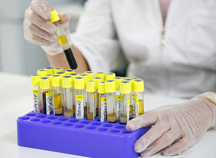
To confirm the diagnosis of dicroceliasis in cattle, research is being carried out in several directions at once. First of all, an analysis of the clinical signs of the disease is carried out. If the result is positive, a laboratory test is performed.
As a material for diagnosis, fresh cow feces are taken. It is examined by the method of successive flushes. The purpose of this analysis is to identify helminth eggs in the feces.

Blood test
You can also determine the infection with parasites on the basis of a blood test. With this type of invasion, an increased amount of immunoglobulin, eosinophils and bilirubin is observed in the blood. This method does not accurately identify the nature of the causative agent of the disease, but may indicate the presence of helminths in the body.
In case of death of the animal, an additional post-mortem examination is carried out before an accurate diagnosis is made. At the same time, the following changes are evidence of the presence of dicroceliasis in livestock:
- traces of exhaustion are clearly visible on the body of the animal;
- the color of the liver is changed to yellow-brown, and the organ itself is noticeably enlarged in size;
- the gallbladder is also enlarged;
- bile ducts inside the liver are dilated and filled with liquid exudate;
- inside the passages, inflamed and dying tissue areas are visible;
- in the section of the liver and gallbladder, a large number of trematodes can be found.
It is worth noting that in order to make an accurate intravital diagnosis, some scientists also advise conducting a study of ants from the grazing areas of the herd. If metacercariae are found in the belly of an insect, then this is another evidence in favor of the presence of dicroceliasis in animals.
Treatment
Treatment of infected livestock is carried out by deworming. It is carried out with the following drugs:
- Albendazole. The composition is used based on the proportion of 0,01 g per kilogram of cow weight. The specified rate of the drug is mixed with animal feed.
- Fenbendazole. It is used in the amount of 33 mg per 1 kg of weight. This anthelmintic is also mixed with food. The drug is fed to the animal once a day for 2 days.
- Fascover. This remedy is used as an intramuscular injection. The dosage is 1 ml per 10 kg of livestock weight. The procedure is carried out once.
- Hexichol. The drug is also mixed with cattle feed. In this case, one dose is 0,3 g per 1 kg of weight. The specified volume of the composition is mixed with 1 kg of compound feed.
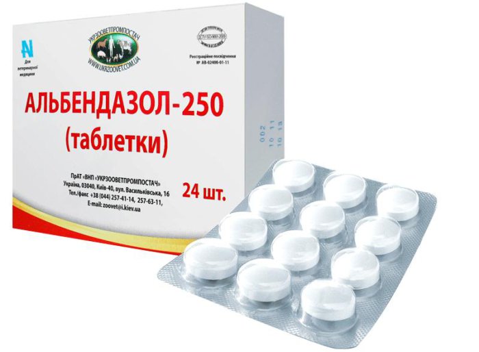
Albendazole
To treat the disease, you can use Hexachlorparaxylol, Bitionol, Fazineks and a number of other anthelmintics. All preparations that are mixed with food can be used in group feeding. If during the course of the disease the animal is greatly weakened, it is better to approach its treatment individually. In this case, intramuscular injections and oral agents are suitable.
Attention! Compliance with optimal conditions of detention significantly accelerates the recovery of livestock.
Prevention
An effective way to prevent dicroceliasis in livestock is periodic deworming of the entire livestock. Moreover, such a procedure is carried out, as a rule, from November to December, when animals are determined for stall keeping. The procedure is repeated before driving the cows to pasture.
To prevent helminths from entering the body, it is better to graze cattle on pastures that are safe for dicrocelia. In addition, the regional ban on the export of manure masses to the territories allocated for pastures allows to reduce the risk of disease in cows.
Improvement of existing grazing areas is carried out by reducing the population of land mollusks. Such a preventive measure is implemented in the following ways:
- Manual assembly. It is carried out in the summer, when the mollusks wait out the heat on the stems and branches of plants.
- Grazing chickens in the pasture. According to the results of research by domestic scientists, it was found that 300 chickens are capable of reducing the number of snails by more than 5% in an area of 80 hectares in 4 minutes. In the next 20 days, this figure rises to 97%.
- Landscaping. This method involves the removal of shrubs, tussocks, debris, stones from the pasture. In this case, the natural nesting sites of snails are eliminated, which leads to a decrease in their population.
- Chemical processing. It is carried out by spraying special chemical compounds on the pasture. These include drug “D”, potassium chloride, metaldehyde in granules (5%).
Also among the preventive measures to combat helminths include regular inspections of the livestock by a veterinarian. Moreover, in the case of importation of livestock from other farms, the animal is placed under quarantine and, in addition to a veterinary examination, its feces are sent for laboratory testing.
Laboratory study of feces
It is worth noting that an important point in the prevention of the disease is also the observance of the basic sanitary and veterinary standards for keeping animals.
Dicroceliasis in cattle can cause considerable harm to livestock farming. Such an ailment can slow down the growth of young animals, reduce the milk and meat productivity of livestock, weaken the body of the animal, as a result of which the risk of secondary diseases increases. The situation is further complicated by the mild severity of symptoms. Therefore, it is much easier to provide livestock with high-quality prevention than to treat a progressive disease in the future.


