Bovine viral diarrhea is a dangerous disease that mainly affects young animals. The disease causes severe disturbances in the work of the intestines in the animal, which are accompanied by diarrhea and a number of other negative consequences. Moreover, if the treatment is not implemented on time, in most cases the disease ends in death.
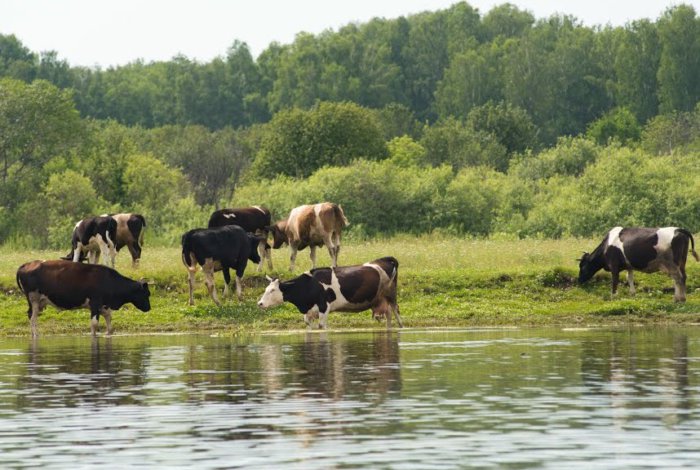
Cattle
What is bovine viral diarrhea?
Viral diarrhea of cattle is an infectious disease that is accompanied by inflammation of the tissues of the intestinal epithelium and abomasum. In this case, the disease also affects the conjunctiva, bronchi, oral and nasal cavities of the animal. In pregnant cows, infection is accompanied by abortion and mummification of the fetus.
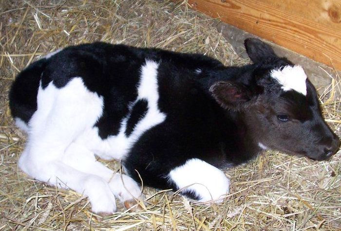
The infection is most often observed in cows aged 6 months to 2 years. During this period, the disease quickly depletes the body of the animal, slowing down its growth and development. In this case, lethality can range from 10 to 90%.
Today, the disease occurs in almost all countries of the world. Due to the high infectivity, the disease manifests itself both in isolated cases and large-scale epidemics. In addition to cows, sheep and goats are also affected by the infection.
Pathogen and its properties
The causative agent of this infectious disease is a special virus from the Pestivirus family. The virion has a spherical shape and dimensions not exceeding 50 microns. Once in the digestive system, the pathogen does not settle in individual organs or tissues, but spreads through the circulatory system throughout the body. In this case, the highest concentration of the pathogen can be traced in the intestines, mucous membranes of the respiratory organs and in the abomasum. The pathogen, while in the body, is able to suppress the immune system, which complicates the fight against it.
The diarrhea virus is also quite resistant to external negative factors. Its pathogenic properties are preserved under the following conditions:
- when heated to 56 degrees, the pathogen survives for 35 minutes;
- at 35 degrees, the pathogen is active for 3 days;
- in tissues and blood, the virus remains viable for several years at a temperature of -20 and below;
- in the body of a cow, the pathogen lives for 120-200 days (with a latent form of the disease and in recovered animals).
The most effective means of pathogen inactivation are boiling, as well as exposure to ether or chloroform. But many researchers argue that individual strains of the virus with high resistance to ether have already appeared today.
The disease can be transmitted through contaminated water
The main source of infection is infected individuals. The virus is released into the environment with milk, feces, saliva, nasal secretions and semen. In healthy individuals, the disease is transmitted in the following ways:
- through feed and water, into which particles of infected feces have fallen;
- airborne droplets;
- in utero from mother to fetus;
- when breastfeeding;
- during mating;
- through the clothes and tools of service personnel;
- through blood-sucking insects and rodents;
- from wild animals.
It should be noted that ill animals can also act as a source of infection. If during the course of the disease the virus was localized in the liver and spleen, then even after complete recovery for another 4 months it remains in the body and can be excreted.
Viral diarrhea appears throughout the year and does not imply a clear seasonality. But in winter, when the body’s immune mechanisms are noticeably weakened, larger epidemics can be traced. At this time, the disease is able to spread to the entire livestock of the farm. In the summer, there are mostly isolated outbreaks of the disease.
Diagnostics
Viral diarrhea in its clinical manifestations is similar to a number of other viral diseases that occur in cattle at calf age. In addition, it is also often confused with bacterial diarrhea. Therefore, for an accurate diagnosis, a comprehensive examination of a sick animal is necessary, including an analysis of clinical signs, pathoanatomical changes, and laboratory research.

Laboratory research
Clinical signs
The incubation period of the virus lasts from 6 to 14 days. At its end, viral diarrhea in cattle can manifest itself in several forms. Accordingly, each of them assumes its own degree of severity of the main symptoms.
The acute form develops mainly in young calves. It is accompanied by such manifestations:
- a sharp increase in temperature to 40-42 degrees;
- strong oppression;
- more frequent shallow breathing;
- accelerated heart rate;
- coughing;
- damage to the mucous membrane of the oral cavity with numerous ulcers;
- outflow from the nasal cavity of exudate with an admixture of pus;
- lacrimation;
- feces are extremely liquid, often with an admixture of blood.
Attention! In some severe cases, the animal may develop conjunctivitis. In addition, frothy secretions from the nose and mouth can often dry out on the animal’s face, forming crusts. Over time, traces of erosion also appear under such crusts.
In the absence of timely therapeutic measures, the main clinical picture is supplemented by a sharp weight loss (up to 25%), dehydration. A rash appears on the genitals, udder and muzzle, which eventually turns into ulcers. The oppression during this period intensifies even more, and the cow almost completely ignores any external stimuli. Due to severe exhaustion, the animal falls into a coma, from which it usually does not come out.
The subacute form of the disease is a consequence of the disease of the representatives of the herd, who have already managed to form a strong immunity. Symptoms in this case are less pronounced. These include:
- a sudden increase in temperature by 2-3 degrees;
- loss of appetite;
- slight oppression of the animal;
- inflammatory and erosive processes on the mucous membranes are less pronounced;
- cough and mucous discharge from the nose may be present;
- lameness, which is the result of damage to cartilage in the joints;
- diarrhea, the duration of which is not more than 24 hours;
Light oppression of the animal
In isolated cases, there is also a sharp reduction in the milk productivity of livestock and cyanosis of the mucous membranes. Calves with this type of diarrhea are slightly behind their peers in weight.
With an abortive (atypical) form of the disease, the symptoms are practically not expressed or may appear periodically. As a rule, the disease in this case passes by itself within a few days, without causing serious consequences.
The chronic variant of the disease is the most difficult to diagnose. In this case, clinical signs develop much more slowly than in the acute form. The disease lasts for six months or more. During this time, diarrhea may occur intermittently or continuously. The calf constantly loses weight, and partial loss of appetite may occur. A lethal outcome in this form of the disease can be traced in most cases.
Pathological changes
When examining a dead animal, the following changes in its body can be detected:
- pronounced effects of dehydration and exhaustion;
- pinpoint ulcers on the mucous membranes, as well as around the nose, on the lips, tongue and abomasum;
- swelling of the tissues of internal organs;
- the liver is significantly enlarged and yellow in color;
- lymph nodes are enlarged, and their internal structure is disturbed;
- the size of the kidneys exceeds the norm, while around them there is a kind of capsule made of fiber, resembling jelly in appearance;
- point erosion of the intestines and blood vessels;
- in the intestines a large amount of exudate with a pungent odor.
It is worth noting that such changes in the internal organs are accompanied by a number of other diseases of the digestive system. Therefore, to confirm the analysis, an additional laboratory study is carried out.
Laboratory analysis
As a material for a detailed study, skin scrapings in places of erosion, blood and washings from the mucous membrane of infected cows are used. If the animal has died, particles of the liver, intestines, spleen and lymph nodes are also taken for analysis.
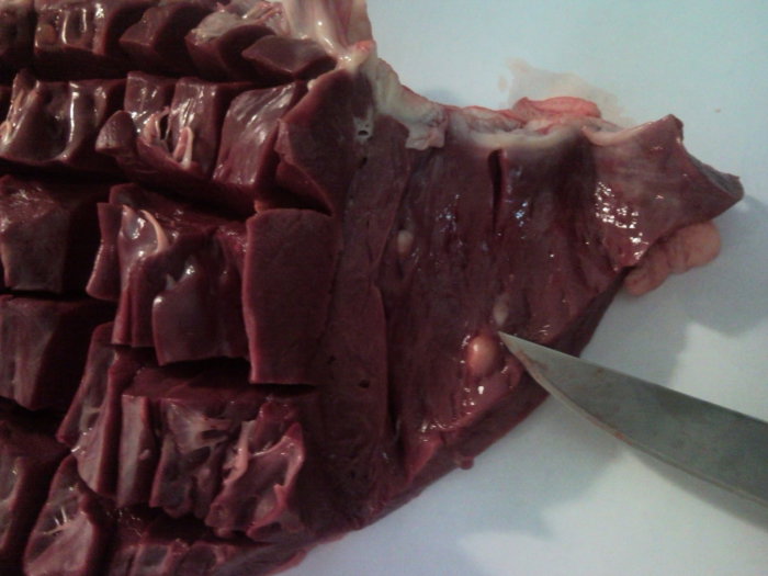
Collection of liver particles for analysis
The resulting material is examined in two different ways:
- Identification of the pathogen and cultivation of a pure culture in an appropriate nutrient medium.
- Detection of antibodies in blood serum by complement fixation or other methods. If the amount of antibodies in the blood exceeds the normal level by 4 times or more, then the diagnosis of viral diarrhea is positive.
Diagnosis of viral diarrhea must be carried out differentially, excluding plague, foot and mouth disease, infectious rhinotracheitis, and stomatitis during the study. All of these diseases are similar to each other in some manifestations, which complicates the diagnosis.
Treatment
The basis for the treatment of such an ailment in cattle is the blood serum of cows that have successfully recovered from viral diarrhea. This substance contains an increased amount of antibodies that can successfully fight the virus. The drug can be administered to livestock in the form of subcutaneous and intravenous injections. But the drug is much more effective in the form of an aerosol. It allows not only to better influence the pathogen, but also to process all the infected livestock at once.
The procedure is carried out according to the following scheme:
- All cattle requiring treatment are driven into a separate sealed box.
- In the same room, a fog generator filled with serum is installed.
- The device is turned on and the drug is sprayed for 40-50 minutes.
- After processing, all cows are returned to their original stalls.
With the development of viral diarrhea, there is a high risk that other infections will enter the body through ulcers that have opened in the intestines and respiratory organs. Therefore, to combat the secondary microflora, various antibiotics are used, among which are:
- Streptomycin. Injections are carried out into the muscles or directly into the abdominal cavity (in the second case, only in combination with novocaine). The standard dosage is from 3 to 10 IU. The introduction is done once.
- Synthomycin. This drug is mixed with livestock feed at the rate of 0,03 g of antibiotic per kilogram of cow weight. The course of treatment is 5 days.
- Levomycetin. It is used in the form of intramuscular injections. The injections are carried out twice a day for 3-4 days. One dose is 10 thousand units.

Injections intramuscularly
Also, instead of antibiotics or in parallel with them, sulfa drugs can be used. They also effectively cope with a bacterial infection.
In addition to the main anti-infective measures, symptomatic treatment of livestock is also carried out. To get rid of indigestion, astringents are used. In this case, tannin and metronidazole are suitable.
In the case of the development of conjunctivitis, the eyes are thoroughly washed with furatsilin, after which they are lubricated with Tilozil, Mezofen or other ointments. The optimal effect is also given by special eye drops.
As for ulcers, it is recommended to wash them with a weak solution of potassium permanganate (potassium permanganate) or furacilin. After thorough disinfection, the affected areas are lubricated with a copious amount of ichthyol ointment.
Often you can also find recommendations for the use of immunostimulants for the treatment of the disease. But they should be used with extreme caution, as in some cases they can only aggravate the situation.
Important! If the animal is in serious condition, intravenous injections of glucose solution (5%) may be necessary to maintain the body. If the virus has also affected the work of the heart, the introduction of adrenaline or caffeine into the blood can be used to stimulate it.
Prevention
Measures to prevent viral diarrhea in cattle are divided into general and specific. General preventive points include the following items:
- organization of balanced nutrition and maintenance of cattle;
- carrying out planned disinfection of premises and care items for cows;
- installation of disinfectant mats at the entrance to the barns;
- mandatory isolation of newly arrived cows for a period of at least 30 days, followed by serological tests;
An effective means of prevention is also the regular treatment of the barn with iodine preparations or a solution of acetic acid. They are good at killing infections. Moreover, it is better to carry out the processing with special fog generators, and do not take the cattle out of the barn for the duration of the procedure.

fog generator
In case of detection of diseased individuals, they are transferred to a quarantine room and individual care and treatment is organized for the animals. At the same time, a number of restrictions are imposed on the economy, which includes a ban on:
- export and import of animals from the farm;
- sale and export of fodder stocks;
- sales of dairy and meat products;
- removal of livestock corpses outside the farm (they are burned right at the place of death).
It is worth noting that although there are special vaccines against viral diarrhea, they are used extremely rarely. General vaccination against infections of the intestines and respiratory tract is more often carried out.
If during quarantine the treatment does not give the desired effect and the animal dies, the room in which it was kept and care items are thoroughly disinfected. To do this, use a solution of phenol or lye.
Viral diarrhea can cause enormous economic damage to farms. It spreads rapidly and has a high mortality rate for young animals. Therefore, in order to prevent the development of the disease, one should strictly adhere to veterinary standards for keeping and feeding livestock, as well as regularly carry out preventive measures. If the animal is still sick, treatment should be started as soon as possible.


