Infectious bovine rhinotracheitis in disadvantaged farms is quite common. At the same time, the disease is accompanied by serious economic losses for the farm, a sharp decline in the productivity of the herd, and the death of animals, especially young animals. Therefore, every breeder must know the main manifestations of the developing disease and how to prevent it.
Infectious bovine rhinotracheitis
What is a disease?
Infectious rhinotracheitis is a disease that affects the respiratory tract and genitals of cows, and can also spread to the central nervous system. In the absence of prompt measures, the infection is quite capable of causing death. Most often, the disease proceeds in an acute form. Young calves under the age of one year are especially susceptible to infection.
Rhinotracheitis (Rinotracheitis infectiosa bovinus) was first identified in 1950. It was discovered by the American scientist Miller while examining livestock. After 4 years, the disease with the same signs was diagnosed in the examined herds by researchers from the USA, Schroeder and Moyes. They also studied the nature of the disease. But only in 1955 the disease was finally studied and received its official name.
Today, rhinotracheitis is widespread in the USA, Africa, Australia, and most European countries. Its causative agent is a special virus, which, according to its external features and properties, belongs to herpes viruses. Infection occurs when it enters the mucous membrane of the respiratory tract or vulva, as well as in the tissues of the prepuce of the penis. In some cases, the infection can be additionally localized in the conjunctiva of the eye, in the tonsils, or even in the brain.
It is worth noting that such a virus is extremely resistant to negative conditions of a harmful environment, which further contributes to its spread. Under the influence of direct sunlight, the pathogen dies within two days. Caustic sodium is able to kill the infection in half a minute. Ethyl alcohol and chloroform destroy the microorganism instantly. But, at a room temperature of 22 degrees, the pathogen is able to live indoors for 50 days. Sub-zero temperatures, up to extreme ones, have practically no effect on the virus.
Reference. When an infection enters the herd, the first manifestations of the disease can be traced in approximately 2-3 weeks. Infection of the entire livestock in the absence of prompt measures occurs after another 3 weeks.
Ways of distribution
The main source of the spread of the infectious rhinotracheitis virus is an infected animal. The disease can affect cattle of any age, regardless of breed. In this case, infection is carried out in the following ways:
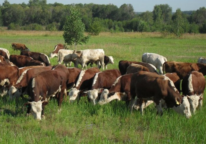
The disease spreads when the herd is heaped up.
- When sick and healthy cows come into contact. It spreads especially quickly when the herd is kept in a heap.
- Respiratory.
- Through the stool. During the development of the disease, the pathogen can accumulate in the intestine, from where it enters the external environment. When such particles enter water or feed, healthy cows become infected.
- Sexually. When the disease is localized in the reproductive organs, the infection is transmitted during insemination.
- Through livestock maintenance equipment, vehicles, staff clothing.
- In birds, rodents and insects. Moreover, the disease may not develop in the carrier, but when it enters the body of cattle, it becomes active again.
Not so long ago, it was found that the virus can also be transmitted through ticks, which animals bring from pastures. Also, the pathogen is able to enter the body of livestock through infected stalls, pastures, and persist for a long time in the grass on pastures.
The most favorable time for the development of the disease is the period from mid-autumn to mid-spring. But even in the summer, cases of the disease are not uncommon.
Pathogenesis
When settling on the mucous membranes, the virus gradually penetrates into their cells and begins to actively multiply. As a result, the affected tissues become inflamed, and after a while they begin to die. Part of the infection enters the blood. Here, the pathogen attaches to leukocytes and, together with the bloodstream, spreads throughout the body. If it enters the conjunctiva, eye diseases additionally develop, including panophthalmitis and conjunctivitis.
If the blood brings the pathogen to the brain, there is a high probability of developing meningoencephalitis in it. This disease is dangerous due to a number of serious consequences, including death. In addition, if a pregnant cow has been infected, the development of the virus in the body ends with the death of the fetus.
Evidence
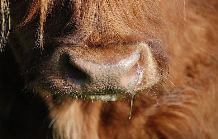
When the pathogen enters the body of an animal, an incubation period passes. It can last from 3 days to a week. At the end of this period, the cow’s temperature rises sharply, which can reach 42 degrees. The behavior of the animal clearly shows a decrease in activity and general depression. In most cases, the development of the disease in the initial stage is also accompanied by a strong cough, heavy breathing, serous viscous discharge from the nose.
Symptom of the disease
All subsequent symptoms of rhinotracheitis can differ significantly depending on the form of the disease, the location of the infection, and the presence of complications. Depending on these factors, the following types of disease are distinguished:
- respiratory;
- conjunctival;
- meningoesophalitis;
- genital;
- atypical.
Respiratory form
It manifests itself acutely, most often develops in young animals. As a rule, respiratory rhinotracheitis is accompanied by the following symptoms:
- The temperature does not fall below 40 degrees.
- Discharges from the nasal cavity become even more abundant and from serous pass into purulent. They may contain blood particles.
- In parallel, intensive salivation develops.
- The developing inflammatory process in the mucous membrane of the respiratory tract is accompanied by the release of a large amount of exudate. It accumulates in the throat and nasal cavity. As a result, the animal begins to breathe more often, shortness of breath quickly appears during movement.
- There is a strong cough.
- On the mucous membrane of the nose, and subsequently the throat, miniature pinpoint ulcers are formed, which develop against the background of tissue necrosis.
- The behavior of the animal is dominated by a sharp decrease in activity, constant lying, and a decrease in appetite.
- Milk productivity drops rapidly and lactation stops completely within 1-2 days.
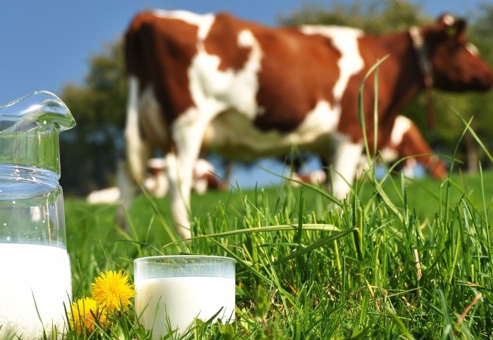
Milk production is falling rapidly
With this type of disease, the infection spreads aerogenically very quickly. Within 7-10 days, it develops in almost the entire herd. The lethality of this form of the disease is about 10%. At the same time, death in an animal occurs from bronchopneumonia, which develops against the background of respiratory rhinotracheitis.
conjunctival form
This type of disease involves the entry of the virus into the mucous membrane of the eye, where it begins to actively develop. As a rule, it gets there already from the mucous membrane of the respiratory tract. As a result, the above symptoms are also added:
- inflammation of the conjunctiva and the formation of edema on it;
- severe redness of the skin around the eye, due to excessive blood supply;
- serous discharge from the mucous membrane of the eye, which can turn into purulent if secondary pathogenic microflora develops against the background of the action of the virus. Such secretions may dry out just below the eyes, forming dry, gray or yellowish scabs.
Reference. The conjunctival variant of the disease of the disease lasts from 3-4 days to 3 weeks.
Meningoencephalitis rhinotracheitis
This type of disease is characterized by the least number of clinical signs. It develops due to the fact that the infection through the blood enters the brain tissue, inflaming its membrane. This form occurs quite rarely and mainly in calves up to 6 months.
Body temperature during meningoencephalitic course of rhinotracheitis practically does not change. Cough, a sharp decline in strength, serous discharge are also absent. The only characteristic signs by which the disease can be identified are severe arousal and problems with coordination in the animal. Also, due to damage to the central nervous system, paralysis of the limbs can develop. The main form of the disease can be supplemented by conjunctival. Death usually occurs within 4 days of the onset of the first symptoms.
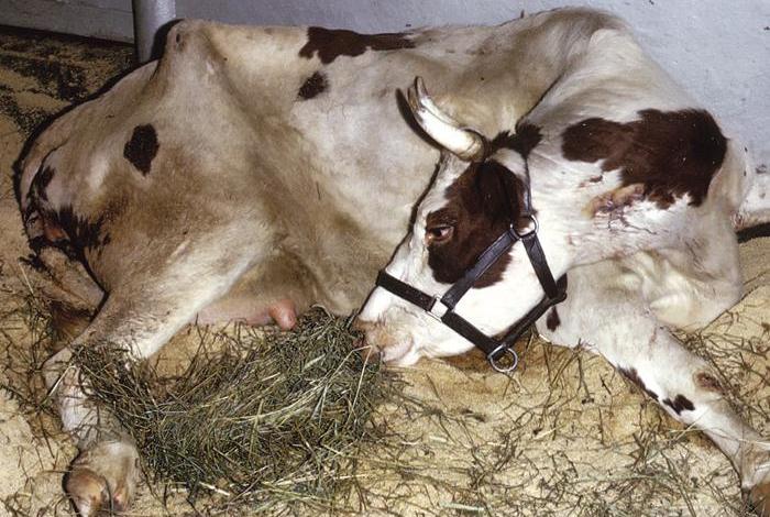
Paralysis of the limbs
genital variety
In this case, the main localization of the pathogen is concentrated in the vulva in cows and on the mucous membrane of the prepuce in bulls. Developing in the external genital organs, the virus causes the development of pustular vulvovaginitis in females, and balanoposthitis in bulls.
In the first case, the following signs are traced:
- decrease in lactation volumes;
- decreased appetite;
- increased blood circulation under the skin in the vaginal area, which gradually turns into edema;
- the vulva is very swollen;
- the cow cannot stand still, constantly stepping over with its hind legs and trying to arch its back;
- ulcers appear on the mucous membrane of the genitals, which are covered with a dark film on top.
With balanoposthitis, the symptoms are:
- Puffiness and swelling of the tissues of the prepuce.
- Pronounced pain during urination.
- In the urine, blood particles can be traced, which are the result of internal hemorrhages.
- After some time, small pustules appear on the tissues of the prepuce, turning into ulcers.
Tellingly, this type of rhinotracheitis can occur subclinically. The symptoms are practically not expressed. Latent flow is especially dangerous for pregnant cows. Within 3-5 weeks, the infection from the genital mucosa through the uterus enters the fetus, as a result of which it quickly dies. Within the next 10 days after this, an abortion occurs.
Atypical rhinotracheitis
This form may be accompanied by most of the symptoms of the respiratory variety of the disease, manifest only by conjunctivitis, or have no clinical signs at all. Often, during its course, accumulation of air in the subcutaneous layer of fiber on the abdomen and thighs can be traced. In some cases, there may be disturbances in the work of the intestine. When a pregnant animal becomes ill, the development of the disease often leads to the death of the fetus.
Diagnostics
According to its clinical manifestations, infectious rhinotracheitis is similar to many other respiratory diseases. Therefore, it is not possible to accurately diagnose a virus developing in the body on the basis of a general picture and a pathoanatomical study. The only way to clearly identify the pathogen is to conduct a laboratory study.

Conducting a laboratory study
This form of diagnosis involves the study of materials taken from a sick animal. In this case, the virus can be detected in two main ways:
- Search for the pathogen itself in tissues and secretions taken from the body. For this, methods of immunofluorescence, enzyme immunoassay are used. Also, due to the fact that the virus is DNA-containing, a DNA probe is often used to identify it, designed to search for a specific nucleic acid of the virus.
- Retrospective study. In this case, the work is aimed at identifying not the virus itself, but the antibodies that are produced in the blood to fight it. For this, the blood serum of a sick cow is used, which is taken twice with an interval of 3 weeks. If, as a result of a serological reaction, 3-4 times more antibodies are found in the second serum, then this particular virus is the cause of pathological changes.
Discharge from the nose, eyes, genitals of the animal can serve as a material for research. If the animal dies as a result of the disease, particles of the mucous membrane of the bronchi, lungs, nose, pieces of lymph nodes or spleen are taken for research. When examining dead cattle, samples are taken no later than 1 hour after their death.
Important! In a separate order, an allergic test is carried out on the skin of a cow.
Treatment
Treatment of cattle in the diagnosis of infectious rhinotracheitis is carried out in a complex manner. Preparations are selected with a focus on such goals:
- The fight against the causative agent of the disease.
- Prevention of development of secondary diseases and complications.
- Strengthening immunity and returning the cow to an optimal state.
To combat the virus, hyperimmune serum is used, as well as blood serum of animals that have recently had rhinotracheitis. The atypical antibodies contained in it help the animal to more effectively resist the developing virus. Most often, such funds are injected under the skin of an animal in an amount of 2 ml per 1 kg of animal weight. The injection is carried out in several places at a time, for a more intensive distribution of the drug. It can also be introduced into the nasal cavity. In this case, the dosage increases to 2-4 ml of the drug per kilogram of weight. This dose is injected into each nostril.
Sulfa drugs and antibiotics are used to combat pathogens of secondary diseases. They are either injected subcutaneously into the animal, or sprayed into the room for the purpose of general disinfection. Also, solutions of hydrogen peroxide and lactic acid are used as an aerosol in barns. In the form of ointments, antibiotics and sulfa compounds are used to treat the genital form of the disease.
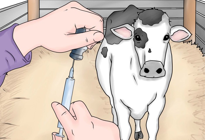
cow injection
In addition to the main course of treatment in order to strengthen the body of livestock, they are provided with balanced feeding with various mineral and vitamin supplements. General strengthening drugs are also prescribed.
Prevention
To prevent the introduction and spread of infection on the farm, basic preventive measures should be strictly adhered to. These include the following points:
- Compliance with basic sanitary conditions for keeping animals. This includes preventing drafts in the barn, adequate insulation, regular cleaning of manure, dirt and leftover feed.
- Proper feeding, preferably individually tailored to the needs of each animal. Ideally, it should be supplemented with vitamin supplements.
- Compliance with quarantine, for a period of at least 30 days, for new animals entering the farm. During this time, the cow is diagnosed and all necessary vaccinations are injected.
- Improving immune mechanisms in the body of livestock. Timely fight against other infectious diseases that can significantly reduce the body’s resistance.
- Periodic disinfection of premises with caustic soda. A hot 4% solution kills the causative agent of rhinotracheitis in a matter of seconds. A 20% bleach solution is also well suited as a disinfectant.
- Immediate isolation of individuals showing clinical signs characteristic of the disease.
Livestock vaccination is also an effective preventive measure. For this purpose, live and inactivated vaccines are used. Among the living, VIEV, “Bivak” and formulations designed to prevent several different infectious diseases at once are especially popular. The vaccine is used for animals at least 10 days old. The injection is carried out twice with an interval of 2 weeks. The resulting immunity is valid for 1 year.
Inactivated vaccines are used exclusively for dairy and breeding farms. Just as in the case of live, inactivated drugs are injected into calves twice. After the second injection, immunity develops within 14 days. Immunity to infection is valid for 12 months.
Conclusion
The main danger of infectious rhinotracheitis is the extremely fast distribution time. At the same time, the consequences of the epidemic of this disease can lead to a significant decrease in the number of livestock on the farm. Therefore, it is important to focus the main efforts on preventing the development of the disease, and when the first clinical signs are detected, immediately contact the veterinary service for help.


