Rectal examination of cows for pregnancy allows you to accurately determine whether the female has been fertilized during mating. You need to know this at the initial stage of pregnancy, because during this special period the animal needs good care and proper living conditions. Such diagnostics makes it possible to identify pathologies in the pelvic organs and other health problems of the cow.
Rectal examination of cows
Research methods
A rectal examination of a cow should be performed by a specialist. This diagnostic method requires careful preparation of both the veterinarian and the animal. The doctor usually works with a partner to help restrain the cow. Research can be done in two ways:
- The animal is standing.
- The animal lies on its left side (used if it is impossible to place a cow).
The cow is tied with a short rope, if necessary, its hind limbs and pelvic region are fixed. Preparation of the veterinarian for the study includes the following steps.
- He thoroughly washes his hands, processes his nails with a file, disinfects the wounds with a disinfectant solution, if they are on the skin.
- The veterinarian puts on high sterile gloves, an oversleeve and an apron.
- The palm is smeared with petroleum jelly or cream for a good glide.
Attention! Rectal diagnostics of a cow is best done in the morning – at this time, animals most often empty their intestines.
The veterinarian starts examining the cow. To do this, he removes the tail to the side and very carefully inserts only his fingers into the anus, while making drilling movements. Next, the doctor pushes them apart so that air enters the anus. Such movement usually provokes the act of defecation.
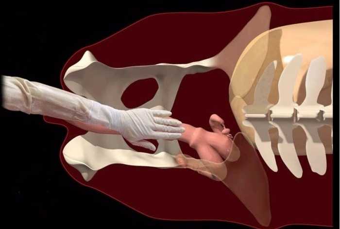
Scheme of rectal examination of cows
The next step is to advance the hand deep into the anus. Soon the hand will rest against the ampulla-shaped extension. After its passage, the intestine narrows. Having penetrated into this section of the intestine, you can begin to study the cow for pregnancy and study its reproductive system as a whole. In close proximity is the uterus and ovaries. To assess the condition of other organs, it is necessary to immerse the hand even deeper.. All movements must be done smoothly and carefully.
Attention! It is unacceptable to make movements with the brush at the moment of muscle contraction – this is fraught with ruptures of the anus.
System Diagnostics
Rectal examination of cows makes it possible to assess the condition of the uterus, its consistency, size, location. But such a diagnosis allows you to examine not only the reproductive system, but also others – excretory and digestive. Having extensive experience, the veterinarian can easily determine the pathologies of various internal organs by their size, density, and tissue consistency. If any abnormalities are found, the animal can be examined more carefully using ultrasound and laboratory tests. Consider how the study of the digestive, excretory and reproductive systems of a cow is carried out by the rectal method.
digestive
When diagnosing the digestive system, the condition of the anus is first assessed. Any deviations are noticeable almost immediately after the introduction of the hand. The diagnostician feels the force of compression of the forearm. Weakness of the sphincter muscles is observed in the following cases:
- In old or debilitated animals.
- In paralysis.
- Dyspepsia.
Reference. An increased tone of the muscle fibers of the sphincter is characteristic of tetanus, and this condition may also indicate blockage of the intestines, displacement or twisting of the intestines.
On palpation, a veterinarian can detect various pathologies of the digestive system – inflammatory processes, intestinal injuries, stretching of the walls of individual sections of the digestive tract, twisting of the loops, their displacement. It is also possible to study the scar by palpation, but this diagnostic method is not considered informative.
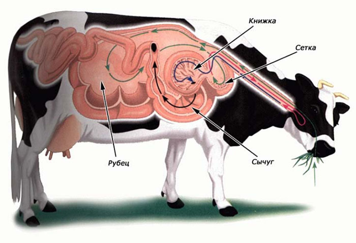
Digestive system of a cow
excretory
With the help of rectal examination, it is possible to assess the state of the excretory system in a cow. In this case, the kidneys and bladder are studied. Diagnosis of the kidneys in too fat animals can be difficult, as these organs are under a dense fatty layer.. In this case, it is difficult for a veterinarian to determine the true size of the kidneys and assess the condition of their surface.
The left kidney is palpated in the region of 3-5 vertebrae in the lumbar region. Normally, it should be movable, while the right one is rigidly attached. By palpation it is possible to evaluate:
- The size of each kidney.
- Examine their surface for tuberosity.
- Define soreness.
The small size of the paired organ indicates a congenital pathology. An increase in the kidneys is most often associated with an inflammatory process. By the presence of irregularities, it can be assumed that the animal is infected with parasites or tuberculosis. If a cow is restless on palpation of the kidneys due to pain, she may have nephritis. Consider how the diagnosis of the bladder is carried out.
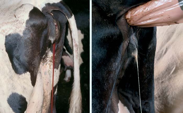
The bladder is located in the pelvic cavity. Its study by palpation allows you to determine the existing pathologies of the excretory system:
- Urolithiasis.
- Paralysis.
- Inflammatory processes in the urinary tract.
- Wall rupture.
- Kidney diseases.
- The presence of a foreign body inside.
More accurate data can be obtained if the study of the bladder in cattle is carried out twice – when the organ is filled with urine and after it has been emptied. Be sure to take into account the tone of the walls of the bladder. Weakness of the muscles of this organ may indicate paralysis if the cow is young.
Sexual
Signs of endometritis
Rectal examination of the reproductive organs makes it easy to determine pregnancy and detect various pathologies of the uterus, cervix and ovaries. Normally, in a non-pregnant cow, the uterus is always in the pelvic region. However, in some diseases, such as endometritis, it descends into the abdominal cavity. The same location is inherent in this organ in the late pregnancy.
The veterinarian should examine the size of the uterus, its density, consistency, degree of fullness, examine its horns. During pregnancy, the uterus has the following characteristics:
- She is enlarged;
- Elastic, soft;
- Easily stretchable;
- The fetus is well felt through its walls.
The condition of the horns also determines whether a cow is pregnant or not. In the third month of bearing a calf, the furrow between the horns is greatly smoothed out.
After examining the uterus, the veterinarian proceeds to palpate the ovaries. His task is to determine the size of each of them, to study their shape. Careful probing reveals cystic formations, as well as the presence or absence of follicles.
Reference. Increased uterine tone indicates that the cow is not pregnant. When pregnancy occurs, the hormonal background changes, while the uterine tone decreases markedly.
Rectal examination of cows is a diagnostic method that is used very widely. Its advantage is low cost, availability and high information content (subject to the competence of the veterinarian). It allows not only to determine whether pregnancy has occurred, but also to identify various pathologies of internal organs – the kidneys, bladder, intestines, uterus and ovaries.


